Penn FTD Center’s Research MRIs: What to Know?
- Penn FTD Center

- Jul 14, 2025
- 3 min read
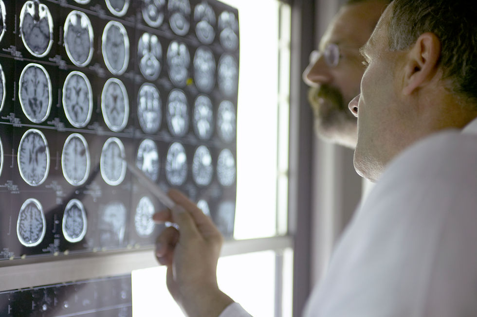
Navigating a neurodegenerative disease is incredibly challenging, and at the Penn FTD Center, we do not want taking part in research to ever be an additional source of confusion or stress for our patients and caregivers. This article highlights some of the key aspects of our research MRIs, how research MRIs differ from clinical MRI scans, and what to expect when coming in for a research MRI.
At the Penn FTD Center, we use two MRI scanners: the Siemens MAGNETOM Prisma (3T) and the Siemens MAGNETOM Terra (7T). The number next to T (Tesla) refers to the strength of the magnet, so a 7T MRI has over two times the magnet strength of the 3T MRI which in turn has two times the magnet strength of a traditional clinical MRI (1.5T). A stronger magnet allows for a higher resolution image that can help us see the structures of the brain more clearly.
Typically, clinical MRIs collect scans to examine neuroanatomy, water content and movement, and blood vessels for diagnostic purposes. In addition to anatomical scans, our research MRIs involve more exploratory sequences that can map connectivity in the brain, quantify iron and myelin content, explore metabolite concentrations through spectroscopy, and so much more, utilizing the physical properties of the brain. These innovative sequences allow researchers to gain vital insights that inform our research work and further our scientific understanding of frontotemporal dementia (FTD) and related disorders.

Our research MRI scanners are in separate buildings from the Perelman Center where the Penn Neurology clinic is located, so when you arrive for a research MRI, you will be accompanied by a coordinator to and from the scanner, and throughout the duration of your research visit. For a 3T MRI, a coordinator will instruct you to prepare for the MRI by removing all metal from your person, including belts, phones, wallets, watches, undergarments that may contain underwire, jewelry, and any additional metal accessories. For a 7T MRI, all participants are also required to change into paper scrubs for an added safety precaution with the higher strength magnet. There are some types of clothing that can actually contain metal fibers, which are undetectable to us as consumers while wearing that clothing, for example, Lululemon Athletica-branded leggings. The paper scrubs are provided to you at the scanner, where there is also a private changing room. Due to this, it is advisable to wear easily removable clothing and shoes to an MRI to reduce the burden of changing clothes.
After you are prepared for the scan, you will enter the MRI control room where an MRI technologist will complete a quick screening with you and then lead you into the scanner room to be positioned on the MRI bed. All research participants are screened at least twice, once with an extensive screening by a research coordinator before the scan is scheduled, and once more with a verbal screening by an MRI technologist prior to the scan.
Our research MRIs last about 60 minutes, so it is recommended that participants use the restroom before entering the scanner. Research participants will have a ball during each scan, which they can squeeze to alert the MRI technologist should they feel uncomfortable at any time, and the technologist will remain in communication with participants throughout the scan.
MRIs are a vital part of our research program at the Penn FTD Center. Participation in research has an enormous impact on advancing clinical care. When the multi-modal data we collect is analyzed together, it allows us to study the biology of FTD and related disorders over time contributing to the improvement of diagnostics, development of new markers of prognosis, and ultimately the discovery of therapeutic targets that can enhance and further the development of treatment trials aimed at treating the underlying biology of FTD and related disorders.
~ Chigozie Ibe
Former Clinical Research Coordinator


